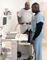
Stress Echocardiogram
What is an Echocardiogram Stress Test?
 An Echocardiogram Stress Test (Stress Echo) is a test that combines an ultrasound study of the heart with a stress test. A stress echo looks at how the heart functions when it is made to work harder. The stress echo is identical to the stress exercise test, except, an echocardiogram is performed before and after you exercise.
The stress echo is especially useful in diagnosing coronary heart disease and the presence of blockages in the coronary arteries (the vessels that supply oxygen-rich blood to the heart muscle).
What does the test show?
An Echocardiogram stress test is performed to evaluate the function of your heart, mainly your left ventricle (main pumping chamber) when the heart is under stress. This test can help evaluate the following:
- Your risk for coronary artery disease.
- If the symptoms you are experiencing (i.e., chest pain or pressure, shortness of breath, unexplained fatigue, palpitations, lightheadedness, etc.) are caused by a blockage to your heart or other heart conditions.
- It can help detect heart problems that may not be present at rest.
- It is used for cardiac clearance before surgery or other procedure.
- If you have already been diagnosed with coronary heart disease, a stress test may enable the doctor to estimate the severity of the blockages.
- If you have just undergone balloon angioplasty or bypass surgery, a stress test can help monitor the success of the procedure as well as determine an appropriate rehabilitation program for you.
Normally, all areas of the heart muscle pump more vigorously during exercise. If an area of the heart muscle does not pump as it should with exercise, this often indicates that it is not receiving enough blood because of a blocked or narrowed artery. The Stress Echo shows areas of the heart muscle that do not receive an adequate blood supply. However, it does not provide images of the actual coronary arteries.
How do I prepare for the test?
- Do not eat or drink for 2 hours prior to the test. This will help prevent the possibility of nausea and vomiting which may accompany vigorous exercise after eating. If you are diabetic or need to eat/drink with your medication, get special instructions from your doctor.
- Avoid any strenuous physical activity on the day of the test because you will need to exert yourself maximally.
- No smoking 2 hours prior to the test. Smoking may interfere with the test results.
- Wear loose and comfortable clothing and shoes that are suitable for exercise; women will wear a hospital gown and men will be asked to exercise bare-chested.
- Do not wear oils or lotions before your test. Small sticky patches (electrodes) will need to stick to your chest.
- Take your medications as prescribed unless your doctor has given you special instructions.
What happens during the test?
When you enter the stress testing room, the Cardiology Tech/Nurse will have you sign a consent form and he/she will make sure you understand the test. Women will be asked to change into a gown and men will be asked to take off their shirt. Your skin will be cleaned to remove any oils or lotions on your skin. You will be shaven if you have a hairy chest. Ten patches are placed on your chest and torso. A belt with wires will be attached to the patches in order to hook you up to the EKG machine. The EKG allows the doctors and Cardiology Tech/Nurse to monitor your heart rate and rhythm. The Cardio Tech/Nurse will take your resting blood pressure and EKG while you are lying down and while you are standing.
The Echo Tech obtains the resting images of your heart while you lie on a hospital table. Gel is applied to the chest and a transducer (small probe) is moved to various areas to obtain the pictures of your heart. The transducer sends ultrasound waves that bounce off the various parts of the heart. These echoes are converted into moving images of the heart. The image is displayed on a screen and recorded on videotape.
The Cardiologist will enter the room before you begin exercising. When the Cardiologist enters the room, he/she will perform a quick assessment, review your medical history, and look at the echo images.
The exercise portion of the test is done on a treadmill. While you are walking, the speed and the grade of the treadmill will increase every 3 minutes. Your blood pressure, EKG and heart rate will be monitored continuously throughout the test. The length of the test varies from patient to patient. However, most patients walk between 6-12 minutes. The treadmill test will stop when:
- You get too tired to continue
- You exceed a "target" heart rate based on your age
- The Cardiologist or Cardio Tech/Nurse detects abnormal changes on your EKG
- You experience symptoms, such as shortness of breath, chest pain, chest tightness, dizziness, etc. that do not permit you to exercise any longer.
- Your blood pressure goes up too high
After the exercise portion of the test, you will be helped to a stretcher. It is important that you get onto the stretcher quickly. The Echo Tech needs to obtain the stress images of your heart while your heart rate is still high. The Cardiologist and Echo Tech compare the two sets of images (before and after exercise) side by side to see how your heart responds to exercise. Your blood pressure and EKG will be monitored during the recovery period.
When do I get the results and what do they mean?
The Cardiologist conducting the test may be able to give you preliminary test results before you leave the testing room. A test report will be sent to your primary Physician in about 3-5 business days. These test results can be discussed during a future office visit.
If your test is positive (abnormal), the Cardiologist conducting the test, along with your Physician, will help develop a treatment plan that is best for you. The Cardiologist may recommend another stress test or more invasive testing such as a cardiac catheterization.
If you have a negative test (no abnormalities) it is likely that your risk of coronary artery disease is low. Stress Echo tests are able to detect individuals with heart disease about 70% of the time. This means that, if you actually DO have heart disease, the test will accurately detect it seven out of ten times.
It should be noted that the Stress Echo test is not 100% reliable. Sometimes the results are "falsely positive" meaning that there is actually no risk of heart disease despite the test's positive results. False positive results occur more frequently in women. Further testing will be necessary to determine whether you actually have heart disease.
If you are concerned about the validity of the test, you may wish to discuss it with your doctor at greater length. You will not be diagnosed with coronary artery disease simply from the results of a Stress Echo test.
Is the test safe?
The echocardiogram stress test is generally safe. There are risks involved because it stresses the heart. Possible rare complications include inducing an abnormal heart rhythm or causing a heart attack.
Where is the test performed?
In the Non-Invasive Cardiology Testing Center and the cardiac rehab facility or in your doctor’s office.
How long does this test take?
Approximately 30 - 45 minutes
Surgeries
- Abdominal Aortic Aneurysm Repair
- Bypass Surgery
- Carotid Endarterectomy (CEA)
- Coronary Artery Bypass Surgery (CABG)
- Transmyocardial Revascularization (TMR)
- Valve Repair Surgery
- Valve Replacement Surgery
Procedures
- Ablation
- Angiojet Thrombectomy
- Aortagram
- Atherectomy
- Automatic Implantable Cardioverter Defibrillators (AICD or ICD)
- Coil Embolization
- Computed Axial Tomography (CAT or CT)/Ultrafact Computed Tomography (CT) Scan
- Coronary Balloon Angioplasty & Stenting
- Coronary Catheterization
- Dobutamine Stress Echo
- Echocardiography (ECHO)
- Electrocardiogram (EKG/ECG)
- Electrophysiology Study (EPS)
- Event Recorder
- Holter Monitoring
- Inferior Vena Cava (IVC) Umbrella Placement
- Intraaortic Balloon Pump
- Intracardiac Ultrasound (ICE)
- Intravascular Ultrasound (ICE)
- Magnetic Resonance Imaging (MRI)/ Magnetic Resonance Angiography (MRA)
- Medicated Stents
- Nuclear Stress Tests
- Pacemakers
- Percutaneous Transluminal Angioplasty (PTA)
- Percutaneous Transluminal Coronary Angioplasty (PTCA)
- Peripheral Stents
- Peripheral Vascular Angiography
- Radiation Brachytherapy
- Septal Closures
- Signal Averaged Electrocardiogram (SAECG)
- Stents
- Stress Echocardiogram
- Stress Test
- Thrombolytic Treatment
- Tilt Table
- Transesophageal Echocardiogram (TEE)
- Valvuloplasty
